Figure 4
From: The role of FGF-2 and BMP-2 in regulation of gene induction, cell proliferation and mineralization

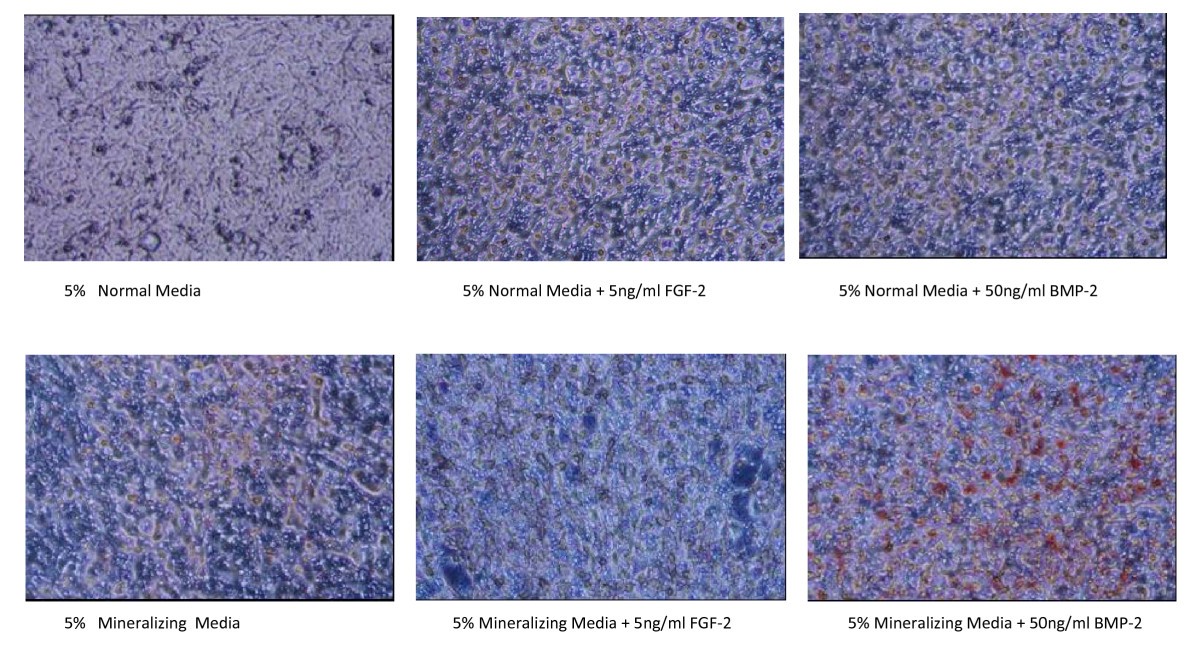
Alizarin Staining of Mineralizing Osteoblast cells. MC3T3-E1 osteoblasts were seeded at 3000 cells/well in 96 well CELLBIND® plates in normal medium. Once cells were confluent, media was changed to 5% NM or 5% mineralizing media with or without 5 ng/ml FGF-2 or 50 ng/ml BMP-2. Two days after treatment, media was removed and cells were fixed in 10% formalin and stored at 4°C until subsequent analysis. Cells were stained for calcium with 2% Alizarin Red for 10 minutes and visualized under 20× objectives for photography. Many areas of mineralization, as seen by bright red staining, were present in the cells treated with 5% MM plus 50 ng/ml BMP-2 (FIG. 11). Little to no mineralization was seen with other 5 treatments.
